


Dark Gums: When Is Pigmentation Natural and When Should It Be Examined?
1. Introduction: Understanding Dark Gums
A healthy smile is not defined by white teeth alone. The colour, health, and appearance of the gums also contribute to an attractive smile. While many people associate healthy gums with a coral-pink shade, gums can naturally range from light pink to brown or dark brown. These variations are often influenced by melanin, the natural pigment responsible for the colour of the skin, hair, and eyes.
Dark gums are not necessarily a sign of poor oral hygiene or an underlying dental problem. In many individuals, darker pigmentation is a normal, harmless characteristic that has been present for years. However, a change in gum colour, the appearance of a new dark patch, or pigmentation accompanied by other symptoms may require professional assessment.
Understanding the difference between natural pigmentation and abnormal discolouration can help individuals make informed decisions about their oral health. This article explores the common causes of dark gums, when they should be examined by a dentist, and the treatment options available when pigmentation requires attention or cosmetic management.
2. Why Do Some People Have Naturally Dark Gums?
The colour of the gums varies from person to person, just as skin colour does. One of the primary reasons for naturally darker gums is melanin pigmentation.
Melanin is produced by specialised cells called melanocytes, which are present in the gum tissue and contribute to its natural colour. When melanin is deposited in greater amounts, the gums may appear brown, dark brown, or nearly black. A dentist in Delhi can examine gum pigmentation and help determine whether it is a natural variation or requires further evaluation.
This condition is commonly known as physiological gingival pigmentation. It is generally a normal variation rather than a disease.
What does natural gum pigmentation look like?
Natural pigmentation may appear in several ways:
- Uniform pigmentation: The gums have a relatively even brown or dark-brown colour.
- Patchy pigmentation: Darker areas appear in certain parts of the gums, particularly around the front teeth.
- Pigmentation along the gumline: A darker band may be visible near the teeth.
- Long-standing discolouration: The gum colour has remained relatively stable over many years.
Natural pigmentation can occur in people of all skin tones, although its appearance and intensity vary. It may affect the upper gums, lower gums, or both.
If the pigmentation has been present for a long time, is stable and is not associated with concerning symptoms, it may simply reflect the individual’s natural gum colour. Nevertheless, appearance alone cannot confirm the cause of every dark patch, so a dental examination is appropriate when the pigmentation is new or uncertain.
3. What Causes Dark Gums Apart from Natural Pigmentation?
Although melanin is a common explanation for darker gums, it is not the only possible cause. Gum discolouration can also occur due to lifestyle habits viz.. smoking , dental materials, certain medications, inflammation, or other conditions.
A. Smoking and tobacco use
Smoking is associated with a condition known as smoker’s melanosis. Exposure to tobacco smoke can stimulate melanin production in the oral tissues, resulting in brown or dark pigmentation, often affecting the front gums.
The extent of pigmentation varies according to individual susceptibility and tobacco exposure. Pigmentation related to smoking may not disappear immediately after a person stops smoking, and any unexplained or changing lesion should be assessed by a dental professional. If gum health concerns are present, periodontics gum disease treatment in South Delhi can help address underlying periodontal conditions based on an appropriate clinical evaluation.
Quitting tobacco is important for overall oral health and can reduce several risks associated with gum disease and oral cancer.
B. Gum inflammation and periodontal disease
Gingivitis and periodontal disease primarily affect the health of the gums and supporting tissues. Typical signs include redness, swelling, tenderness, and bleeding during brushing or flossing.
These conditions do not always cause dark pigmentation directly. However, inflammation, bleeding, healing changes, and deposits around the teeth can alter the appearance of the gumline. If dark areas appear alongside persistent bleeding, swelling, or discomfort, a dentist should examine the gums to identify the underlying cause.
Professional cleaning and appropriate periodontal treatment may be recommended when plaque, tartar, or gum disease is present.
C. Certain medications
Some medications can cause pigmentation of the oral tissues as a side effect. The likelihood depends on the specific medicine, dosage, duration of use, and individual response.
A dentist may ask about current and previous medications when investigating unexplained gum discolouration. Patients should not stop prescribed medication independently; any suspected medication-related pigmentation should be discussed with the prescribing doctor.
D. Dental materials and previous dental treatment
Occasionally, dark or blue-grey areas near a tooth may be associated with dental materials. For example, an amalgam tattoo can develop when small particles from an older silver-coloured filling or dental procedure become embedded in the surrounding soft tissue.
This may appear as a flat, localised blue-grey or dark patch. Although such marks are often harmless, a new or unexplained pigmented area should not automatically be assumed to be an amalgam tattoo without appropriate clinical assessment.
E. Injury or local trauma
Minor trauma to the gums can sometimes cause bruising or a temporary change in colour. The affected area may appear dark red, purple, or almost black, depending on the nature of the injury.
The expected course depends on the cause and severity. If the discolouration persists, enlarges, recurs, or is accompanied by pain or swelling, it should be assessed rather than attributed to trauma without confirmation.
F. Less common medical or oral conditions
Certain systemic conditions and uncommon oral lesions can also cause changes in pigmentation. These possibilities are particularly important when discolouration develops suddenly, spreads, appears different from the surrounding tissue, or occurs with other symptoms.
Most dark gum pigmentation is not cancer. However, a dentist must consider the appearance, history, and clinical characteristics of an unexplained lesion before deciding whether further investigation is necessary.
4. When Is Dark Gum Pigmentation Natural, and When Should It Be Examined?
One of the most important questions is whether the gum colour has always been present or whether it represents a recent change.
Long-standing, stable pigmentation without other symptoms is more consistent with a natural variation. In contrast, new or changing pigmentation deserves attention because its cause may not be immediately obvious.
Signs that may be consistent with natural pigmentation
The following features can be reassuring, although they cannot establish a diagnosis on their own:
- The darker gum colour has been present for years.
- The pigmentation has remained relatively unchanged.
- The colour is distributed in a pattern consistent with the person’s natural pigmentation.
- There is no associated pain, ulceration, unexplained bleeding, or swelling.
- Routine dental examinations have not identified any concerning changes.
Warning signs that should not be ignored
Arrange a dental assessment if you notice:
- A new dark spot or patch on the gums.
- A dark area that is increasing in size or changing in colour or shape.
- An irregular or unusually coloured patch that differs from the surrounding tissue.
- Pigmentation associated with an ulcer, lump, swelling, pain, or unexplained bleeding.
- A persistent sore or other oral lesion that does not heal.
- Numbness or other unexplained changes in the affected area.
A persistent mouth sore, red or white patch, lump, or unexplained oral bleeding warrants prompt assessment, particularly if it lasts more than two weeks. Do not wait for two weeks if a lesion is rapidly changing or otherwise concerning.
These symptoms do not necessarily mean cancer is present. Many oral conditions are non-cancerous, but early professional evaluation helps ensure that significant conditions are not overlooked.
Important: A painless dark patch is not automatically harmless. Some concerning oral lesions may initially cause few or no symptoms. A dentist may recommend further investigation or referral to an oral medicine specialist or oral surgeon if the appearance is suspicious.
5. How Does a Dentist Examine Dark Gums?
A dental examination helps distinguish likely physiological pigmentation from discolouration that needs further investigation.
Step 1: Medical and dental history
The dentist will ask when the pigmentation first appeared, whether it has changed, and whether there are any associated symptoms. Relevant details may include tobacco use, medication history, previous dental treatment, medical conditions, and any history of trauma.
Step 2: Clinical examination
The dentist will inspect the gums and surrounding oral tissues, assessing the colour, distribution, size, shape, and surface of the pigmented area. The surrounding teeth, gumline, cheeks, tongue, and other relevant oral tissues may also be examined.
The examination helps determine whether the pigmentation appears consistent with a normal variation or whether further evaluation is necessary.
Step 3: Additional investigation, if indicated
Many cases of typical, stable physiological pigmentation can be assessed clinically without special tests.
If the cause remains uncertain or the lesion has suspicious features, the dentist may recommend specialist evaluation and, where appropriate, a biopsy. A biopsy involves taking a small tissue sample for microscopic examination to help establish a diagnosis.
Dental X-rays are not routinely required simply to assess surface gum pigmentation. Imaging may be used if there is a separate clinical reason to investigate the teeth or underlying bone.
The appropriate approach depends on the individual findings rather than the colour of the gums alone.
6. What Treatment Options Are Available for Dark Gums?
Treatment depends on the cause. Natural gum pigmentation does not usually require treatment for health reasons. If the pigmentation is a cosmetic concern, however, a dentist can discuss suitable options after confirming that the tissue is healthy.
A. Treating the underlying cause
If the discolouration is related to a dental or medical condition, management focuses on the cause rather than simply removing the pigment.
Depending on the findings, this may involve professional cleaning, treatment of gum disease, addressing local irritation, or coordinating with the patient’s doctor regarding suspected medication-related changes.
Suspicious lesions require appropriate diagnostic investigation before any cosmetic procedure is considered.
B. Gum depigmentation for cosmetic concerns
Gum depigmentation is a procedure intended to reduce excess visible pigmentation in selected areas of the gums. It may be considered by people who feel self-conscious about darker gums, particularly when the pigmentation is visible while smiling.
One technique involves using a dental laser to remove or reduce the superficial pigmented gum tissue. Other techniques may be suitable in selected cases, depending on the clinical situation and the dentist’s assessment.
At Smile Delhi – The Dental Clinic, laser gum depigmentation using a Biolase laser is an available cosmetic treatment option for suitable cases. Many patients from across the globe have benefited from this procedure in Smile Delhi-The Dental Clinic .
The procedure is not necessary for everyone with dark gums, and it should only be considered after a thorough examination.
C. What should patients know before gum depigmentation?
Before proceeding, patients should understand that:
- The procedure is cosmetic when performed for natural pigmentation.
- Suitability depends on the health of the gums and the pattern of pigmentation.
- Healing time varies according to the technique, treated area, and individual response.
- Temporary soreness or sensitivity may occur during healing.
- Pigmentation can recur because pigment-producing cells may remain or become active again.
- Tobacco use may contribute to recurrent pigmentation and can adversely affect oral health.
The dentist will explain the expected healing process, aftercare instructions, potential risks, and realistic outcomes before treatment.
Gum depigmentation should never be used as a substitute for investigating an unexplained or suspicious dark lesion.
7. Can Dark Gums Be Prevented?
Natural melanin pigmentation cannot necessarily be prevented, and darker gums do not indicate that a person has poor oral hygiene.
However, certain habits can help maintain healthy gums and reduce avoidable oral health risks:
Maintain good oral hygiene. Brush twice daily with a fluoride toothpaste and clean between the teeth regularly. This helps control plaque and supports gum health.
Attend routine dental check-ups. Regular examinations allow the dentist to assess changes in the gums and identify potential problems early.
Avoid smoking and tobacco products. Tobacco can affect gum pigmentation and increases the risk of periodontal disease and oral cancer.
Discuss unusual changes promptly. Do not attempt to scrub, bleach, or apply home remedies to dark areas of the gums. Household bleaching agents and unverified products can damage delicate oral tissues.
Follow personalised dental advice. If pigmentation is associated with medication, inflammation, or another condition, follow the recommendations of the appropriate healthcare professional.
Good oral hygiene supports gum health, but it does not necessarily change natural melanin pigmentation.
8. Dentist’s Insight: Why Understanding the Cause Matters
Dark gums are a good example of why dental care should focus on both appearance and health. A darker gum colour may be completely natural, and cosmetic treatment is not automatically necessary. At the same time, a new pigmented lesion should not be dismissed simply because dark gums are common.
The most important factors are the history of the pigmentation, any recent changes, the appearance of the surrounding tissues, and the findings of a clinical examination.
For patients considering cosmetic gum depigmentation, the priority is to confirm that the gums are healthy and that the pigmentation is suitable for treatment. A careful assessment helps distinguish a cosmetic concern from a condition that needs investigation.
The goal is not to make every person’s gums the same colour. It is to maintain healthy oral tissues, recognise unusual changes, and offer appropriate treatment when it is genuinely needed.
9. Conclusion: Should You Be Concerned About Dark Gums?
Dark gums can be a natural and harmless variation caused by melanin, particularly when the pigmentation has been present for years and remains stable. However, a new, changing, irregular, or symptomatic dark patch should be examined by a dental professional.
The right approach begins with understanding the cause. Some cases require no treatment, others may benefit from management of an underlying dental issue, and selected patients may consider gum depigmentation for cosmetic reasons.
At Smile Delhi – The Dental Clinic, New Friends Colony, South Delhi, an individual assessment can help determine whether gum pigmentation is a natural variation, needs further investigation, or may be suitable for cosmetic treatment.
If you have noticed a change in your gum colour or would like to explore options for dark gums, arrange a dental consultation for personalised advice.
We Care To Make You Smile.
Frequently Asked Questions
1. Are naturally dark gums unhealthy?
Not necessarily. Natural melanin pigmentation is common and does not, by itself, indicate gum disease or poor oral hygiene. The overall health of the gums and any changes in their appearance are more important than colour alone.
2. Can dark gums indicate a serious problem?
Sometimes, but most dark gum pigmentation is not cancer. A new, unexplained, growing, irregular, or symptomatic dark patch should be examined by a dentist to determine the cause.
3. Does everyone with dark gums need gum depigmentation?
No. Gum depigmentation is an elective cosmetic procedure for suitable patients. Natural pigmentation generally does not require treatment for health reasons.
4. Is laser gum depigmentation permanent?
Results vary. Pigmentation may return over time because pigment-producing cells can remain or become active again. Your dentist can discuss the likelihood of recurrence based on your individual circumstances.
5. Can smoking make gums darker?
Yes. Smoking can stimulate melanin production in the gums, causing smoker’s melanosis. Quitting tobacco is beneficial for oral health, although existing pigmentation may not disappear immediately.
6. Should I wait if a dark patch does not hurt?
No. The absence of pain does not establish that a lesion is harmless. Arrange a dental examination for any new or changing dark patch, especially if it has an unusual appearance or is accompanied by other symptoms.
Sources
- American Dental Association — Oral Cancer Guideline — evaluation and referral of suspicious oral lesions.
- National Institute of Dental and Craniofacial Research — Oral Cancer — warning signs and the importance of early examination.
- Colgate India — Causes of Black Gums — general information on natural melanin pigmentation and changes in gum colour
Posted by Dr. Sonakshi

Dr. Suprriya B. Bhatia is a highly experienced dental professional with over 25 years of clinical expertise. A leading dentist in South Delhi, she is the founder of Smile Delhi – The Dental Clinic and specialises in cosmetic dentistry, dental implants, and smile makeovers. Dr. Suprriya B. Bhatia is known for her compassionate, patient-centred approach and commitment to creating healthy, confident smiles for patients from India and abroad.


"Extremely good service and care..."
Tim Gound
Location:- U. K.


"The standard of the clinic is like a first country’s clinic..."
PEDRO MORAES
Location:- Brazil


"Me and my family were very reluctant to undergo my dental treatment in India, but..."
RICHARD SALDANHA
Location:- Peru, South America


"The hygiene is good, the care up to European standards if not better. Strong on explaining..."
THOMAS BAINS
Location:- European Commission

"A new beautiful smile made by a beautiful lady who truly cares for her patients..."
Sinead
Location:- England

"I have the smile of my dream….This clinic is very professional and they use only best materials..."
Alla Lu
Location:- Russia


"Thank you so much for making my dream come true to have a perfect smile..."
Jacyln Gopinathan
Location:- Mindro Philippines


"I felt very comfortable through my treatment and i had 100% trust in you..."
Arnar Gislason
Location:- Iceland


"Thank you for giving me a smile that i’ve dreamed of for years..."
Yuto Harrison
Location:- Canada


"Dr. Suprriya’s work was amazing…. She is super professional and polite..."
Todd
Location:- Australia


"Dr. Suprriya is such a lovely and warm hearted woman as well as a truly and high qualified professional..."
Victor Vique
Location:- Switzerland


"After travelling all through India and visiting 7 different clinics, this..."
JEFF MC CLEAN
Location:- New Zealand

"From the moment i got here i knew i had made the right decision."


"Thank you so much, Dr. Suprriya! I am so grateful for you and your practice."

"Very attentive and grateful for the results. I will keep in Touch"

"You saved my teeth. Lovely clinic and very professional staff..."


"My teeth are 7-8 shades whiter, thanks to you and your team..."

"Thank you very much dr. suprriya for wonderful treatment for implants and bridge and giving me my smile back."


"Felt in good hands during the whole treatment..."

"Visited many dentist around the world and smile delhi definitely gives you a great smile..."


"Over all experience was wonderful…"
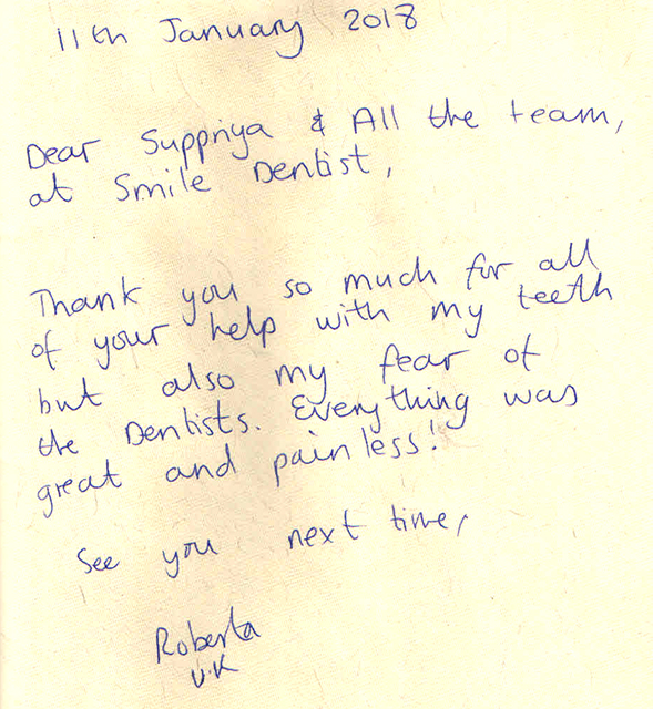
"Every thing was great and pain less !....."
Roberta
Location:- UK

"Dr. Suprriya is such a lovely and warm hearted woman as well as a..."
Victor
Location:- Switzerland


"Didn’t hurt, looked good, felt good, clearly explained and professionally done..."
Jeff
Location:- New zealand

"Dr. Supria is a unic dentice and an exellent person..”, “…honest and direct.."
Yaron Mayer
Location:- Israel Embassy

"Dr. Suprriya is very gentle and excellent human being. A perfect Dentist I...."
Uarely
Location:- Finland


"You treated me gentle as a child!..."
Andreas
Location:- Norway


"You are probably one of the best dentist I have ever been to in the last 50 years..."
Usama
Location:- Saudi Arabia


"I have the Hollywood teeth I always wanted…"
Belle
Location:- Canada

"The root canal was almost pain free....."
David Seeley
Location:- UK

"Thank You very much for giving me my smile back and my holiday!....."
Lesley Marsdin
Location:- UK

"I even laughed when they screwed the posts in ! very professional even enjoyed it a bit"
Laurewce Moon
Location:- UK

"It has been very professional. Very clean, very good......"
Mr. & Mrs. Piper
Location:-Winchester, England

"My emergency treatment was incredible kind and 1st class....."
Kevin Kahan
Location:- Ireland
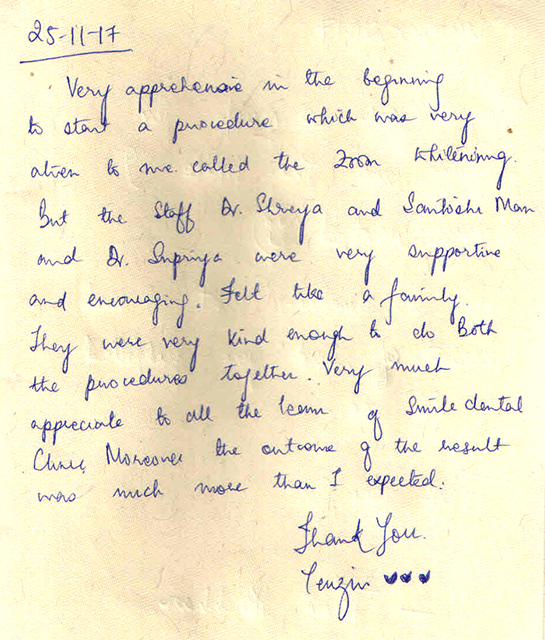

"The result was much more than I expected in zoom whitening....."
Tenzin
Location:- Bhutan


"Root canal was pain free and i felt really comfortable during the procedure....."
Angie
Location:- Paraguay

"Amazingly professional and courteous experience......"
Ken Bakshi
Location:- USA


"I had my teeth whitened amazing results!"
Paula Davey
Location:- Australia


"I would like to have sometimes so good dentist in Europe like Dr Suprriya..."
DANNY
Location:- Austria

"I would highly recommend Suprriya to anyone seeking good oral health care..."
ALLISON WEST
Location:- Australia


"Dr Suprriya, I just want to thank you for the very well done..."
MARIO
Location:- Columbia (Diplomat in India)